
EXPLORE CELL MECHANICS 1-WELL DYNAMIC CONFINER
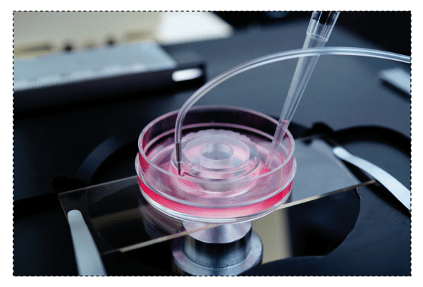
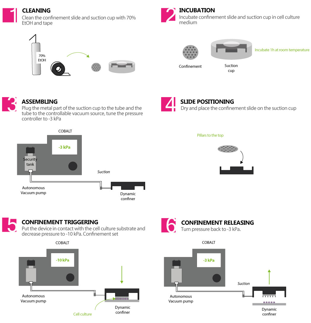
The dynamic cell confiner is a device that confines cells between two parallel surfaces to a defined micrometer height with sub-micrometer precision. The space between the two surfaces is controlled by micro PDMS pillars. The micropillars are fabricated on a glass slide, which is attached to a PDMS piston (suction cup). The piston is controlled with a vacuum pump, and the height of the confinement is thus controlled as well.
> CONTROL THE CONFINEMENT SPEED
Accurate control of the confinement speed through the vacuum pump
> REVERSIBLE CONFINEMENT: RETRIEVE YOUR CELLS AFTER CONFINEMENT
Allows molecular analysis thanks to the non-destructive method for the cells
> COMPATIBLE WITH YOUR OWN EXPERIMENTS
The confiner is a small size device that is directly placed on the top of your cell culture
FEATURES & BENEFITS
THE DYNAMIC CELL CONFINER KIT IS COMPOSED OF:
> 1 x Cobalt autonomous vacuum pump (visit our partner’s product webpage to know more)
> 12 x Confinement Slides – 10 mm (diameter) glass slides/cover slips with micro-structures in PDMS (pillars) that enable the confinement.
Available confinement heights – from 1, 2, 3, … up to 20 µm (choose up to 3 heights to integrate your kit)
> 3 x 4Dcell Dynamic Confiner™ (PDMS Suction Cup)
> 1 x Security tank (to preserve the vacuum pump)
> 1 x Kit of Tubes and Connectors
> 3 x PDMS ring shaped stencil – delimits the seeding area of cells to the region where confinement is applied
IF YOU WANT TO MAKE YOUR SLIDES ADHERE TO YOUR CELLS OR NOT (OPTIONAL):
> Aliquots of extracellular matrix protein for cell adhesion (for example, fibronectin) in the right buffer solution
> Aliquots of anti-adhesive molecule (poly-ethylene glycol) ready to bind on the slides
CANCER
> Migration of metastatic cells
> Cell contractility in mestastasis
> DNA DSB repair (mechanically induced)
> Genomic instability (cell division)
> Separated co-culture
IMMUNOLOGY
> Migration of immune cells
> Imaging of non-adhesive cells
ORGAN PHYSIOLOGY
> Migration of cancer cells
> Cell differenciation with stiffness control
> Wound healing
> Separeted co-culture
> Cell compression response
RARE DISEASES
> Cell nucleus integrity
AGING
> Cell nucleus integrity
> Autophagy related diseases
OBSERVATION OPTIMIZATION
> Imaging of non-adhesive cells
> Planar imaging of organelles
FUNDAMENTAL RESEARCH
> Cell volume (cell cycle)
> Cell stretching response
Video of HeLa cells under confinement using a 4Dcell confiner, going from initial state to extremely confined.
Video of cells dividing under confinement using a 4Dcell confiner
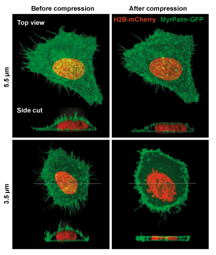
Example of mammalian cells with before and after confinement images with fluorescent proteins
Le Berre M, Aubertin J, Piel M., Fine control of nuclear confinement identifies a threshold deformation leading to lamina rupture and induction of specific genes. Integr Biol (Camb). 2012 Nov;4(11):1406-14.
> A flagellate-to-amoeboid switch in the closest living relatives of animals
Brunet. T., et al., Elife, 2021 Jan 15;10:e61037
> The nucleus measures shape changes for cellular proprioception to control dynamic cell behavior
Venturini. V., et al., Science, 2020, 370, eaba2644
> Confinement and Low Adhesion Induce Fast Amoeboid Migration of Slow Mesenchymal Cells
Y.-J. Liu, M. Piel, et al., Cell, 2015 160(4), 659-672
> Actin flows induce a universal coupling between cell speed and cell persistence
P. Maiuri, R. Voituriez, et al., Cell, 2015 161(2), 374–386
> Geometric friction directs cell migration
M. Le Berre, M. Piel, et al., Physical Review Letter 2013 111, 198101
> Mitotic rounding alters cell geometry to ensure efficient spindle assembly
O. M. Lancaster, B. Baum, et al., Developmental Cell, 2013 25(3), 270-283
> Fine Control of Nuclear Confinement Identifies a Threshold Deformation leading to Lamina Rupture and Induction of Specific Genes
M. Le Berre, J. Aubertin, M. Piel, Integrative Biology, 2012 4 (11), 1406-1414
> Exploring the Function of Cell Shape and Size during Mitosis
C. Cadart, H. K. Matthews, et al., Developmental Cell, 2014 29(2), 159-169
> Methods for Two-Dimensional Cell Confinement
M. Le Berre, M. Piel, et al., 2014, Micropatterning in Cell Biology Part C, Methods in cell biology, 121, 213-29