Endothelial tubulogenesis assay
Improved endothelial cell tubules formation

Improved endothelial cell tubules formation
Polyacrylamide gels; Microstructured polyacrylamide gels; Microchannels
The random distribution of endothelial cells in standard culture conditions do not resemble the blood vessel network typically found in vivo.
3D patterned endothelial cells show improved formation of functional vascular networks and can be employed for instance in tissue engineering. In addition, endothelial cells geometrically organized in 3D microstructures provide a better in vitro model to study tubulogenesis.
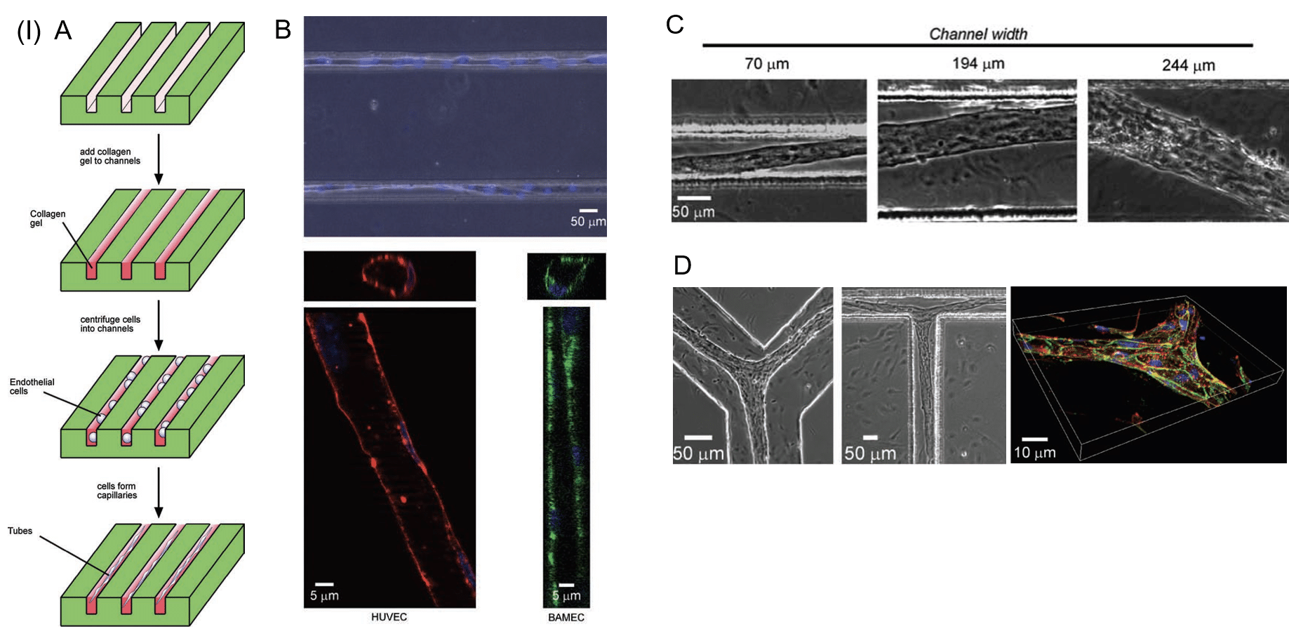
(I) Microfabricated channels induce the formation of functional endothelial cell tubes. A: Schematic representation of endothelial cells within channels containing a matrix of collagen. B: Endothelial cells (24 h post-culture) are spatially organized in collagen-containing microfabricated channels (upper panel – phase contrast; lower panel – fluorescence, red and green stains actin, and blue stains for nuclei). C: Larger channels widths lead to the formation of larger diameter tubes. D: Branched patterned templates enable the formation of bifurcated capillary networks. Cell-cell junctions visible after b-catenin staining (green; actin – red; nuclei – blue). HUVECs, human umbilical vein endothelial cells; BAMECs, bovine adrenal microvascular endothelial cells [1].
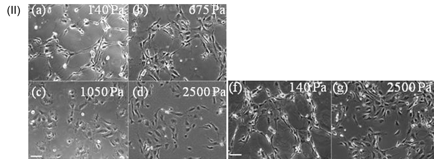
(II) Gel stiffness determines the successful formation of endothelial cell networks. Endothelial cells form networks on gels of low stiffness (140 Pa) whereas in hard gels the cells are not able to assemble into a stable network (2500 Pa) [2].
[1] Srivatsan, R. et al. TISSUE ENGINEERING: Part A Volume 16, Number 7, 2010
[2] Randi L. Saunders and Daniel A. Hammer. Cell Mol Bioeng. 2010 March ; 3(1): 60–67