Lamellipodia and filopodia assay
Analyzing events occuring during membrane protrusion

Analyzing events occuring during membrane protrusion
Actin organization, cell adhesion and polarization
Cells plated on culture dishes lost cell-cell and ECM-cell interaction leading to the loss of their reproducible shape and cytoskeleton organization. In addition, localization of the lamellipodia formation is difficult to observe. Lamellipodia are an actin projection on the leading edge of the cell.
Using 4Dcell squared micropatterns, lamellipodia tend to go at the corner of the cells patterned making easier their behavior study.
Endothelial cells on disk shape compared to squared micropatterns [1].
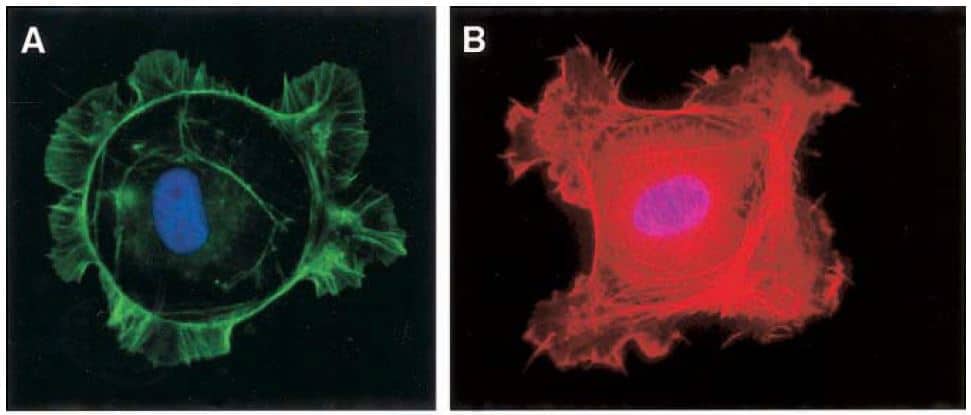
A. In a disk micropattern, cell processes are extended at random points
B. In a square micropattern, cell processes extended from their corners.
Blue: DAPI to visualize nuclei ; Green and red: fluorescent phalloidin to visualize F-Actin with lamellipodia and filopodia.