
5000+ ORGANOIDS
5000+ ORGANOIDS
5000+ ORGANOIDS
CELL CONFINEMENT
CONTROL CELLS BETWEEN TWO PARALLEL SURFACES
PHYSIOLOGICAL ASSAY AT A 2.5D LEVEL
Cell confinement is a powerful approach to recreate the native mechanical and spatial environment of cells, offering a more realistic alternative to traditional 2D cultures.
At 4Dcell, we refer to our advanced confinement technologies as operating at a 2.5D level, a sweet spot between the simplicity of 2D and the complexity of 3D, enabling more physiologically relevant assays without sacrificing experimental control.
Cells in vivo are rarely spread flat on a glass surface. They are surrounded by tissues, experience spatial constraints, and interact with their microenvironment in three dimensions. Through precise geometrical confinement, we guide the shape, polarity, and mechanics of individual cells or small colonies. This enhances the biological fidelity of in vitro experiments, leading to more predictive insights in cell behavior, drug response, and differentiation.
Whether you are studying stem cell fate or cancer cell migration, 2.5D confinement allows you to tune mechanical cues and spatial organization with high reproducibility, critical for reliable and scalable drug discovery workflows.
By mimicking the architecture of real tissues while maintaining accessibility and imaging compatibility, 4Dcell’s confinement systems bring the next level of physiological relevance to your cell culture assays.


Easy to use
High resolution imaging
Long term cell culture
PLATE YOUR CELLS. CONFINE. MEASURE.
CONFINEMENT TECHNOLOGY
Cells are homogeneously confined/compressed between two parallel surfaces to a defined micrometer height with sub-micrometer resolution.
-
Different confinement heights available (e.g. 1 µm – 300 µm)
-
Allows long-term cell culture and cell proliferation to proceed while preserving perfect control of confinement
-
Compatible with high-resolution optical microscopy systems
-
Can process a sufficiently large number of cells to allow whole gene expression analyses to be performed
-
Can be combined with bio-functionalized micro-structured substrates and/or with different matrices (geometry control)
-
Can be combined with gels (stiffness control)
-
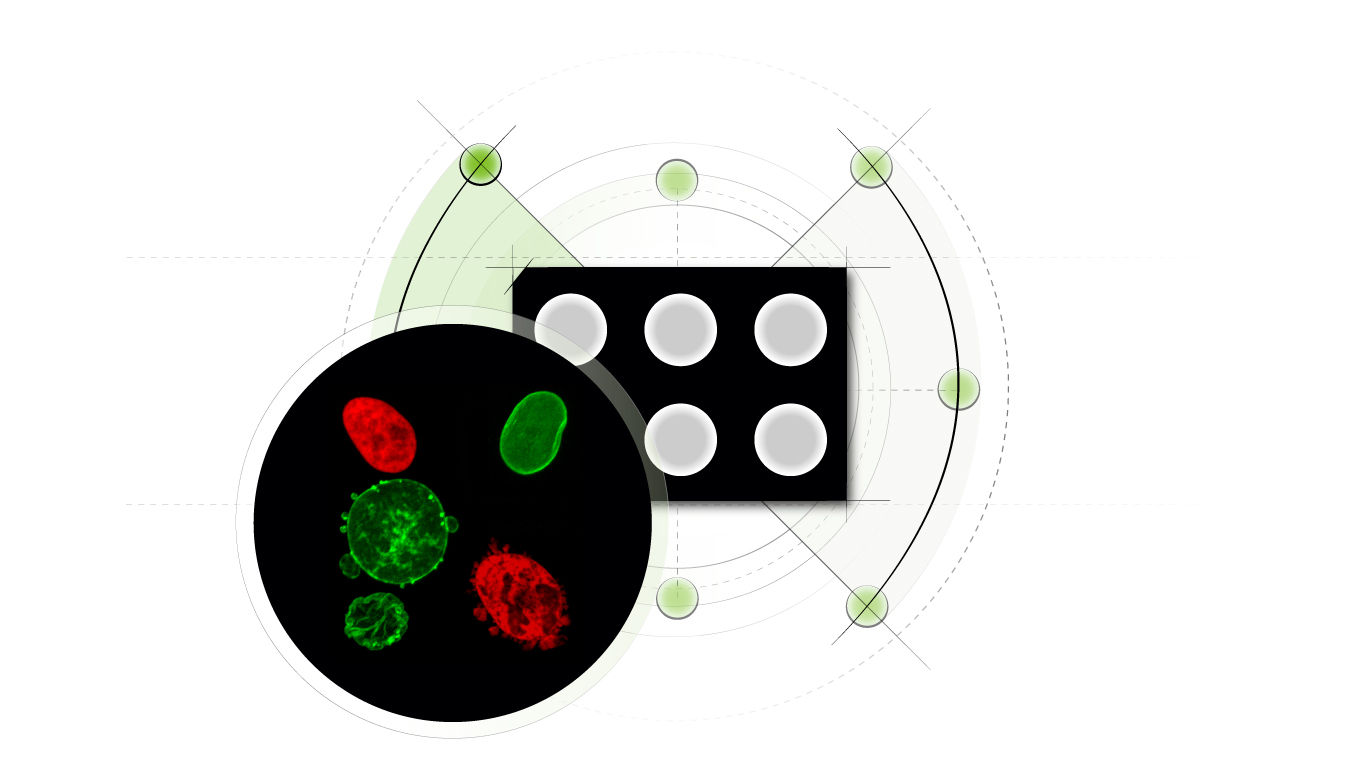
Compatible Petri dishes of 35 mm and 6-well plates
-
For customized designs, please contact us

CONFINEMENT APPLICATIONS
Cell migration 2.5D, migration and interaction of non-adhesive cells, cell squeezing, imaging of flat cells (organelles aligned in 2D), super-resolution video-microscopy (organelles move less), contractility assay, etc


HeLa cells: not confined, 5 µm, 3 µm.
Nuclear bebling and laminar rupture is modulated by the degree of cell confinement
Cell migration phenotype behaviour is affected by degree of confinement height and adhesion
Cells undergo phenotypic transition in response to confinement height
Domains of application
CANCER
-
Migration of metastatic cells
-
Cell contractility in mestastasis
-
DNA DSB repair (mechanically induced)
-
Genomic instability (cell division)
-
Separated co-culture
IMMUNOLOGY
-
Migration of immune cells
-
Imaging of non-adhesive cells
ORGAN PHYSIOLOGY
-
Migration of cancer cells
-
Cell differenciation with stiffness control
-
Wound healing
-
Separeted co-culture
-
Cell compression response
RARE DISEASES
-
Cell nucleus integrity
AGING
-
Cell nucleus integrity
-
Autophagy related diseases
OBSERVATION OPTIMIZATION
-
Imaging of non-adhesive cells
-
Planar imaging of organelles
FUNDAMENTAL RESEARCH
-
Cell volume (cell cycle)
-
Cell stretching response
STUDY CELL DYNAMICS TRIGGERED BY/UNDER MECHANICAL EFFECTS
-
Migration
-
Cell division
-
Induce autophagy
-
Mechanotransduction
-
Mechanics of the nucleus
Explore examples of applications
-
Cancer invasiveness assay: Quantification of migration behaviors and migration transitions
-
Cancer aggressiveness assay: Quantification of contractility of somatic or cancer cell
-
Immune system in a well: 2D migration and interaction of non-adherent immune cells
-
Immune cells interaction: 2D interaction of non-adherent immune cells
Video of HeLa cells under confinement using a 4Dcell confiner, going from initial state to extremely confined.
Video of cells dividing under confinement using a 4Dcell confiner

Example of mammalian cells with before and after confinement images with fluorescent proteins


