
SmartHeart®
A human-relevant fully integrated cardiac model to identify safer and more effective molecules.
SmartHeart captures what hERG and 2D don't see.

3-in-1 3D cardiac assay replicating key in vivo mechanisms
The SmartHeart is an integrated solution to measure contractility (1), action potential (2), and calcium handling (3).This streamlined approach minimizes variability, delivering unmatched insights into in vivo toxic mechanisms
CONTRACTILITY (1)

Contractility Metrics
Frequency
Contraction stress
Contraction strain
Contraction speed
Relaxation speed
Drug cardiac tropism
Chronotropic
Inotropic
Chronotropic
Lusitropic
ELECTROPHYSIOLOGY (2)

Action potential Metrics
Frequency
Interpeak duration STD Rising time, Upstroke velocity
APD10-90
Triangulation, Plateau phase
Electrophysiology features
Rhythm
Regularity Depolarization
Impacted channels Proarrhythmia
CALCIUM TRANSIENTS (3)

CaT Metrics
Frequency
Interpeak duration STD Amplitude
Area Under curve ‘Rise and decay’ duration and speeds
CD10,50,90
Comprehensive understanding of calcium dynamics in tissues


Brightfield - Contractility

FluoVolt - membrane action potential

CAL520 - Calcium transient
UNPRECEDENT FEATURES

HUMAN HEART TISSUES
hiPSC-CMs and cardiac fibroblast

HIGH THROUGHPUT
96-well plate

FULLY INTEGRATED MODEL
Coupled contractility, electrophysiology, and calcium readouts

SELF-ASSEMBLY / EASY TO USE +++
Plate your cells, let them beat, measure

7200 CELLS:TISSUE
3:1 ratio of cardiomyocytes to fibroblasts

HIGH STATISTICAL POWER
9 replicates per well

HIGH RESOLUTION IMAGING
Glass bottom plate

IN SITU CHARACTERIZATION
Multiple key readouts in a single assay
SMARTHEART CAPITALIZE ON THE HIGHEST STANDARDS
A human-like cardiac model engineered to reach a high level of maturation
SmartHeart® tissues offer enhanced morphological, structural, molecular, and functional maturity, increasing the predictability of cardiotoxicity studies and clearly surpassing the limitations of traditional 2D approaches.

3D reconstruction of a ring. Vimentin (green), Troponin T (red) and DAPI (blue)

Immunostained contractile fibers at 63X magnification. Troponin T (red) and DAPI (blue) – Seguret & al.
MORPHOLOGY
The ring-shaped geometry allows for a more physiologically accurate distribution of forces within the tissue. It facilitates the visualization of re-entrant waves, which are responsible for most clinical arrhythmias.
STRUCTURE
The SmartHeart® platform provides structurally mature tissues featuring well-aligned contractile fibers and sarcomeres.

MOLECULAR EXPRESSION
RT-qPCR data highlight the upregulation of key genes for cardiac function compared to 2D models.
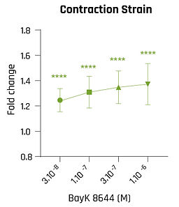
Direct measurement of positive and negative inotropy, what 2D and CHO can't give you
Inotropy, the modulation of cardiac contractile force, is a key efficacy and safety signal that conventional hERG, CHO, and 2D hiPSC-CM assays cannot measure. SmartHeart® quantifies it directly through contraction strain in 3D human cardiac tissue, capturing both positive and negative inotropy with statistical confidence.
Positive inotropes
Negative inotropes


Why it matters: SmartHeart® detects negative inotropy driven at the ion-channel level, mirroring the cardiodepressant effect of dihydropyridine calcium blockers.
Why it matters: The canonical positive-inotropy benchmark confirms a physiological β-adrenergic response with a clean dose-response.
Why it matters: Detects inotropy driven at the ion-channel level, with statistical significance even at the lowest concentration tested

Why it matters: A myotrope that raises the calcium transient, invisible to calcium- or voltage-only assays, but clearly resolved by SmartHeart®'s direct force readout.

Why it matters: Captures calcium-handling-based inotropy, the mechanism of next-generation heart-failure therapeutics.

Why it matters: a receptor-level negative inotrope, confirming that SmartHeart® captures the full bidirectional inotropic spectrum.


PACE X WELL-BY-WELL STIMULATION SYSTEM

Unpaced

1.5 Hz

2 Hz

2.5 Hz
Precision & Consistency of Stimulation
Well-by-well pacing ensures each well receives the exact same stimulus, eliminating variability caused by spontaneous beating or uneven stimulation. This leads to cleaner signals, reduces experimental noise, and minimizes artifacts due to differences in cell behavior or plating, ultimately improving data quality and assay reliability.
Improved assay reproducibility
Standardized pacing minimizes discrepancies between wells, improving intra- and inter-plate reproducibility.
Multiplexing of conditions
Different compounds, cell types, or pacing frequencies can be applied simultaneously in the same plate, ideal for high-throughput screening (HTS).
Synchronization with data acquisition
Timing of contraction events can be aligned with optical or impedance-based readouts, enhancing signal clarity and consistency.
Enhanced model translatability
Regular pacing mimics physiological heart rates, increasing the relevance of the assay for predicting in vivo responses (e.g. inotropic effects, QT prolongation, arrhythmia risk).
Supports cardiomyocyte maturation
Chronic pacing promotes structural and functional maturation of hiPSC-derived cardiomyocytes (sarcomere organization, gene expression, calcium handling).
Enables kinetic or dose-response studies
With consistent pacing, subtle changes in contraction amplitude, duration, or rhythm in response to drugs can be detected with higher sensitivity.
SMARTX 2.0 AI DRIVEN PRECISION FOR YOUR EXPERIMENTS
Bright field videos (e.g. 500x500 pixels, min 35 fps and 10 s length) of the contracting rings can be analyzed using 4Dcell’s SmartExplorer software, SmartX 2.0.
The algorithm uses machine learning to detect and track the area of the central pilar, from which it determines several parameters: contraction amplitude, contraction strain, contraction and relaxation speed, contraction stress, etc.


SmartX is an AI-powered image analysis platform engineered to evolve with every experiment you run.
At its core lies a deep learning module that continually “learns” from each dataset, refining its ability to detect and track beating rings with ever-greater accuracy and speed. Whether you’re measuring contraction strength, frequency, or spatial consistency, SmartX adapts to your unique imaging conditions and cell patterns, automatically adjusting its segmentation models to reduce false positives and enhance signal fidelity.
The result is a self-optimizing workflow that delivers robust, reproducible metrics experiment after experiment, freeing you to focus on biological insights rather than manual analysis.