Cell volume assays
Tracking the cell volume of adherent cells in real time

Tracking the cell volume of adherent cells in real time
Cell volume measurement technology
Cell volume, ion pumps
There are several physiological and pathological processes where cells undergo a change of volume. However, there are no reliable methods that can be applied to accurately measure volume of adherent cells in real time.
Cells are cultured in an optically transparent chamber that enables to accurately determine their volume along time and to follow in parallel the biochemical processes responsible for the volume change, as for example activation of ion pumps.
Volume tracking from interphase stage, to mitosis of Raji cells [2].
(A) Cells are placed in poly(dimethylsiloxan) chambers of calibrated height set by pillars, in medium supplemented with FITC-Dextran. Bottom picture: cells exclude fluorescence on epifluorescence images (Scale bar 20 mm).
(B) The fluorescence profile corresponding to the dotted line in (A): maximum and minimum of fluorescence intensity correspond to chamber maximal height (background) and zero height (pillar), respectively. Right: these values are used to calibrate the signal and calculate the optical thickness of the cells.
(C) Finally, cell volume is obtained by integrating the total fluorescence intensity over the cell area.
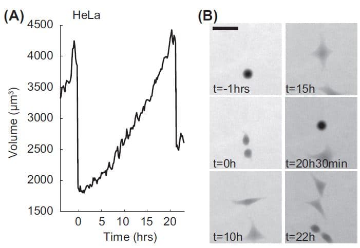
(A) Volume trajectory of a HeLa cell. The two volume overshoots at the beginning and the end correspond to transient volume increase in mitosis with the first one corresponding to the mother cell and the second one to the daughter cell.
(B) Raw Fluorescence images of the cell in (A) with FXm. Scale bar 50 mm.
[1] Zlotek-Zlotkiewicz, E. et al. (2015). Journal of Cell Biology, 211(4), 765–774.
[2] Cadart, C et al. (2017). Methods in Cell Biology, 139,103-120.