SMART[contractility] Plates: Contractile ring shaped organoids
Explore this easy to use technology developed by 4Dcell to measure contractility force

SMART[contractility] Plates: Contractile ring shaped organoids
Explore this easy to use technology developed by 4Dcell to measure contractility force

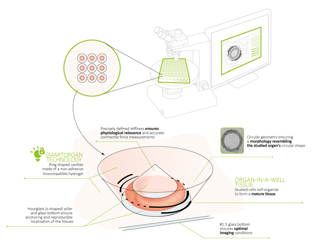
4Dcell SmartContractility plates offer an easy way to self-assemble 3D aggregates of cells into rings that facilitate contractility force measurements. The plates are coated with a 3D structured hydrogel, molded into an array of conical-shaped microwells containing a central pillar. When cells are seeded on top of these microstructures, they are guided towards the ring-shaped cavity, self-assembling into a circular organoid, which surrounds a hydrogel central pillar.
The stiffness of the hydrogel is tuned so that contraction of the ring-shaped organoid results in the deformation of the central pillar. The contractility force is then quantified from the deformation of the central pillar and its known mechanical properties.
> READY TO USE
Plate your cells directly on the microstructured substrates
> KEEP YOUR ORGANOIDS IN PLACE
Due to the innovative design, the rings are kept in place during manipulation
> CONTRALITY FORCE MEASUREMENT
Measure the contractility force through the deformation of the hydrogel based central pillar
> PREDICTABLE LOCATION AND EASY TO IMAGE
Cell aggregates are located in predictable positions enabling automated image acquisition (bright field, fluoresence, phase contrast, etc.)
> TUNABLE STIFFNESS OF THE SUBSTRATE
Control the substrate stiffness mimicking the in vivo environment of your cell type
FEATURES & BENEFITS
WORKING PRINCIPLE OF THE SMART[contractility] TECHNOLOGY AND FEATURES:
Scheme of the working principle of the structured hydrogel responsible for the formation of the ring-shaped organoids.
> The cell culture substrate is based on a biocompatible Polyethylene glycol (PEG) hydrogel with molded microwells containing a central pillar.
> The hydrogel is cell repellent, precluding the adhesion of cells to the substrate.
> The inclined walls (funnel shape) of the microwells allow the cells to be guided towards the circular cavity, thus decreasing the number of cells lost in the seeding process.
> The ring shaped organoids are kept in place during medium exchange, addition of drugs, etc. This is achieved because the bottom of the microwells are made of glass, enabling the organoid to anchor to this area. In addition, the central pillar has an ‘s’ shape profile, preventing the rings from sliding up due to contractions.
> The soft nature of the hydrogel (5 kPa to 20 kPa) enables the quantification of the contractility force through the deformation of the central pillar.
> The gel stiffness can be tuned according to the cell model in place.
> The cell culture substrates are compatible with image acquisition, as the cells are seeded on areas composed of only glass, the PEG hydrogel surrounding the cells is transparent and #1.5 glass (ideal for most objectives) is used.
TECHNICAL SCHEME OF THE SMART[contractility] TECHNOLOGY:
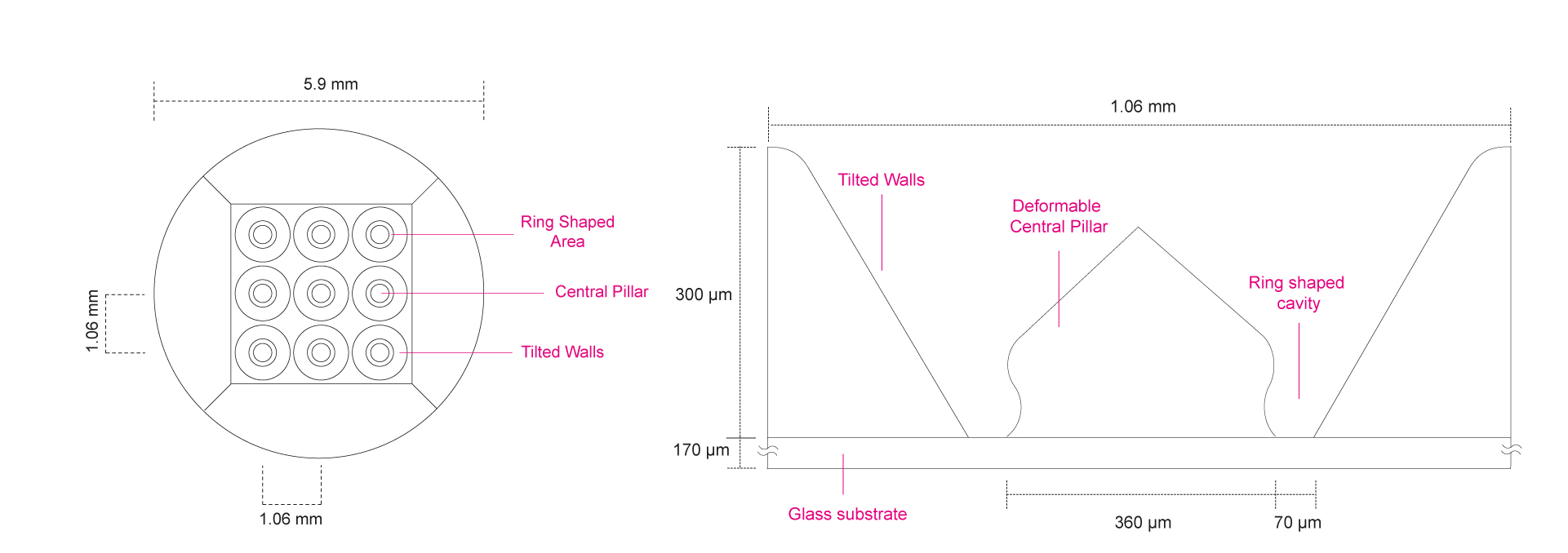
SMART[contractility] PLATES:
6, 12 and 24 well plates with glass bottom ( #1.5, 0.16 – 0.19 mm)
Each well has 9 individual microwells for organoid formation
The gels are provided in an aqueous solution that retains their mechanical and optical properties
CANCER
> Signaling pathways
> Immune activation
> Tissue Structure
DRUG SCREENING
REGENERATIVE MEDICINE
TOXICOLOGY
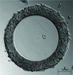
Mouse embryonic fibroblasts, 22 hours after being seeded.
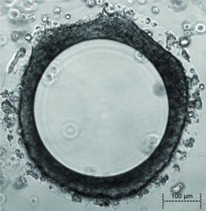
Mouse embryonic fibroblasts, 12 days after being seeded.