Smooth Muscle Cells Assay
Alignment, organization and shape standardization

Alignment, organization and shape standardization
Alignment and organization of smooth muscle cells
In vivo, cells are highly organized to form structures, tissues, and organs – and that structure helps determine function. For example, smooth muscle cell alignment around the small intestine yields a tissue capable of exerting substantial force. Conventional 2D cell culture conditions – in which cells are given a boundless surface on which to grow in any orientation – don’t reflect the cell’s natural microenvironment.
When cultured in a substrate with line adhesive cues, smooth muscle cells get organized and acquire an elongated shape.
4Dcell partnered up with Cell Systems to test the smooth muscle cells assay. Cell Systems is an innovative company that provides cells and strives to give the most biological and physiologically-relevant conditions for research. They have antibody-free primary cells which are ideal tools for optimizing your experiments. Not only is it hard to find a more biologically-relevant cell type, but their deep inventory guarantees access to the same cell population for the entirety of your research program. This means more consistency and increased reproducibility.
ACBRI 716 Primary Human Aortic Smooth Muscle Cells, from Cell Systems cultured on 4Dcell micropatterned slides (Panels A and B) versus standard tissue culture dish (Panel C). Each surface was coated with Attachment FactorTM extracellular matrix and all cultures were maintained in Complete Classic Medium with serum and CultureBoostTM.
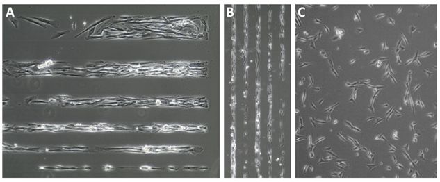
Phase contrast images captured 2hr after plating. Magnification: Panel A, 20X; Panels B and C, 10X.