
EXPLORE CELL MECHANICS 6-WELLS STATIC CONFINER – CSOW 620
The CSOW 620 – static confiner is a portable device that allows confining cells within two surfaces to a defined micrometer height and with nanometer precision. The space between the two surfaces is controlled by using micro PDMS pillars. The micro pillars are fabricated in a glass slide, which is attached to a PDMS piston.
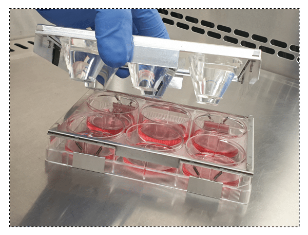
The CSOW 620 is the device that controls the position of the pistons and it is compatible with glass bottom Falcon 6-well plates. The system is designed for long term confinement of cells. In the end of the confinement experiments, the cells can be retrieved enabling further biochemical studies. The user decides when and where to confine the cells as it is not necessary to plug it to any other device.
The CSOW 620 is characterized by its compact design and possibility to be plugged to a CO2 source.
> DEFINE THE THICKNESS AND SHAPE OF CELLS
Control the thickness of your cells with the right confinement slide
> MULTIPLE EXPERIMENTS SIMULTANEOUSLY
Enables the study of different cells or to apply different confinement conditions at the same time
> ADAPTABLE WITH HIGH RESOLUTION MICROSCOPY
Optically transparent materials and compact design enable high resolution microscopy
FEATURES & BENEFITS
THE STATIC CELL CONFINER CSOW 620 IS COMPOSED BY:
> 1 x CSOW 620
> 12 x Confinement Slides – 16 mm (diameter) glass slides/cover slips with micro-structures in PDMS (pillars) that enable the confinement
Available confinement heights – from 1, 2, 3, … up to 20 um (you can choose up to 3 heights to integrate your kit)
> 12 x Confinement Piston in PDMS
IF YOU WANT TO MAKE YOUR SLIDES ADHESIVE TO YOUR CELLS OR NOT (OPTIONAL):
> Aliquots of extracellular matrix protein for cell adhesion (for example, fibronectin) in the right buffer solution
> Aliquots of anti-adhesive molecule (poly-ethylene glycol) ready to bind on the slides
> If this device does not fit with your constraints, get in touch with us! We can personalize it for you
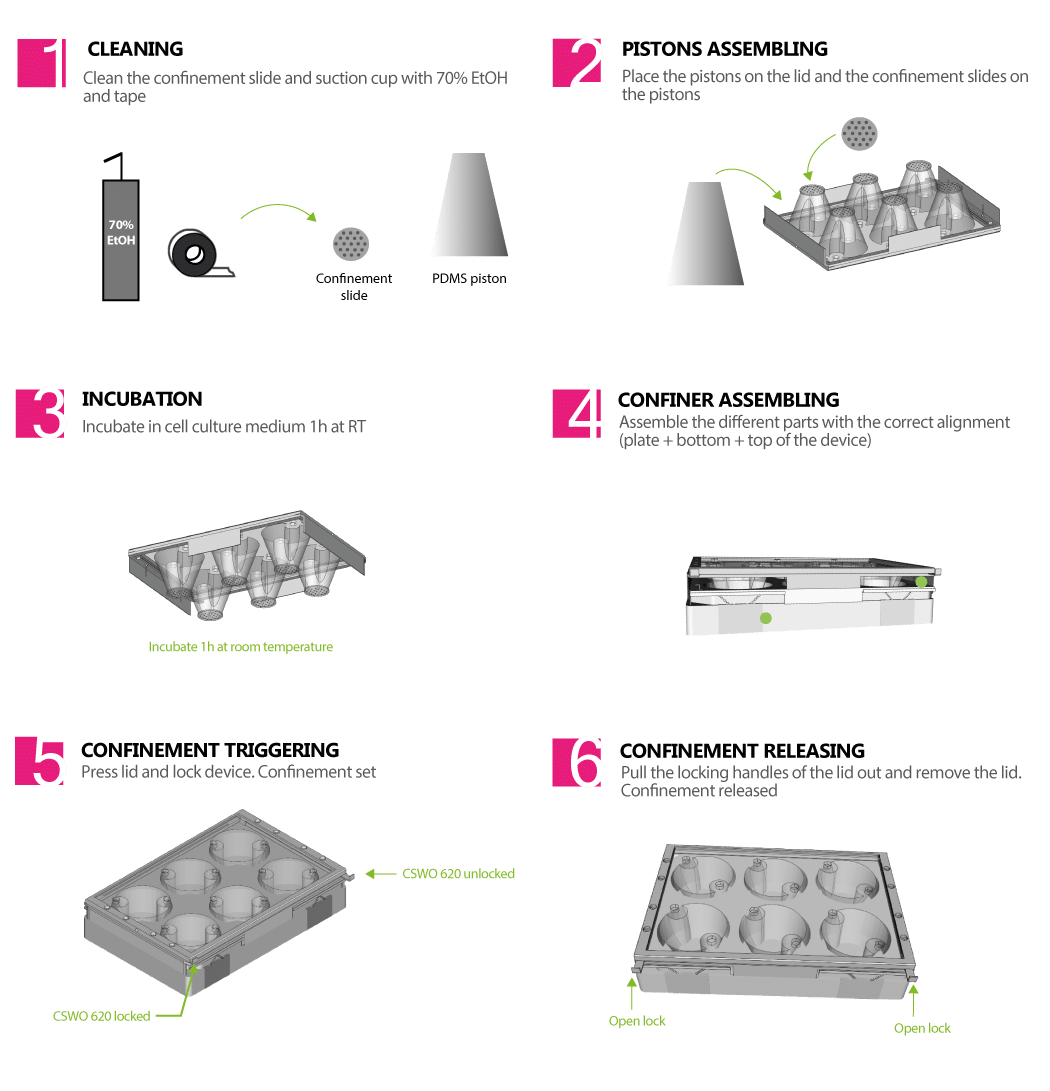
VIDEO PROTOCOL TO SET UP PERFECTLY YOUR CSOW 620
ANATOMY OF THE CSOW 620


Overall dimensions of the CSOW 620
> TO STUDY CELL DYNAMICS TRIGGERED BY/UNDER MECHANICAL EFFECTS
Migration
Cell division
Induce autophagy
Mechanotransduction
Mechanics of the nucleus
> HIGHT RESOLUTION IMAGING
> CO-CULTURE OF CELLS
Video of HeLa cells under confinement using a 4Dcell confiner, going from initial state to extremely confined.
Video of cells dividing under confinement using a 4Dcell confiner
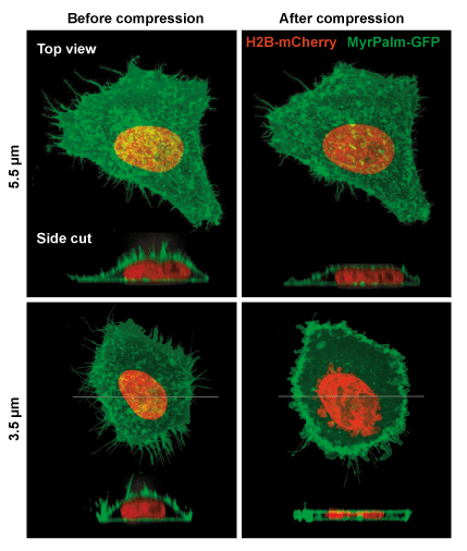
Example of mammalian cells with before and after confinement images with fluorescent proteins
Confinement and Low Adhesion Induce Fast Amoeboid Migration of Slow Mesenchymal Cells
Y.-J. Liu, M. Piel, Cell, et al., 2015 160(4), 659-672
Actin flows induce a universal coupling between cell speed and cell persistence
P. Maiuri, R. Voituriez, et al., Cell, 2015 161(2), 374–386
Geometric friction directs cell migration
M. Le Berre, M. Piel, et al., Physical Review Letter 2013 111, 198101
Mitotic rounding alters cell geometry to ensure efficient spindle assembly
O. M. Lancaster, B. Baum, et al., Developmental Cell, 2013 25(3), 270-283
Fine Control of Nuclear Confinement Identifies a Threshold Deformation leading to Lamina Rupture and Induction of Specific Genes
M. Le Berre, J. Aubertin, M. Piel, Integrative Biology, 2012 4 (11), 1406-1414
Exploring the Function of Cell Shape and Size during Mitosis
C. Cadart, H. K. Matthews, et al., Developmental Cell, 2014 29(2), 159-169
Methods for Two-Dimensional Cell Confinement
M. Le Berre, M. Piel, et al., 2014, Micropatterning in Cell Biology Part C, Methods in cell biology, 121, 213-29