Single cardiomyocyte contractility force assay
Quantification of beating force of single cardiomyocyte cells

Quantification of beating force of single cardiomyocyte cells
Uniaxial Contraction Force, Analysis of Calcium Transients coupled to force measurements.
When hESC-Cardiomyocyes are cultured in standard conditions, i.e., in 2D monolayers of cells, these are organized in a complete random way. Therefore, it is not possible to precisely quantify the uniaxial force generated by the beating of a single cardiomyocyte cell.
In this assay, derived cardiomyocytes attach to rectangular cell-adhesive areas, which induce cell elongation and promote suspended cell anchoring between two adjacent micropillars. The contractility force assay enables in vitro reproduction of excitation−contraction coupling effect responsible for the beating of CMs.
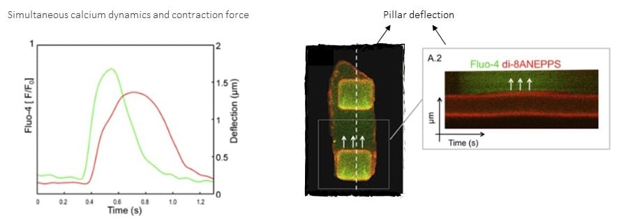
Quantification of calcium transients (activation of calcium-sensitive contractile proteins) and simultaneous measurement of cell contraction and force generation (analysis of pillars deflection) in hESC-Cardiomyocytes [1].