
SMARTHEART®
3D Cardiac Contractility & Inotropy Assay

Quantify positive and negative inotropic drug responses in human 3D cardiac microtissues.
SmartHeart® combines human iPSC-derived cardiomyocytes, cardiac fibroblasts and mechanically loaded, ring-shaped microtissues to quantify changes in contraction, relaxation and beating activity.
Run your experiments in your own laboratory using SmartHeart® 96-well plates, or work with 4Dcell to access our fit-for-purpose qualified cardiac contractility testing service.
The platform supports pharmacological characterization, cardiac efficacy studies and functional safety assessment.
QUANTITATIVE TISSUE-LEVEL CARDIAC CONTRACTILITY
Unlike assays based solely on ion-channel activity or cellular motion, SmartHeart measures mechanical deformation in human 3D cardiac microtissues contracting against a deformable hydrogel pillar.
Brightfield video recordings are analyzed using SmartX 3.0 to quantify 22 morphological and contractile metrics, including:
Inotropy
Contraction strain and derived contractile stress to characterize positive and negative inotropic effects.
Chronotropy
Beating frequency and beat-to-beat irregularity to identify changes in spontaneous cardiac activity.
Contraction & Relaxation Kinetics
Contraction and relaxation speeds and timing parameters to assess changes in mechanical activation and recovery.
Together, these complementary readouts provide a quantitative functional profile of compound-induced cardiac responses.
CHARACTERIZING POSITIVE AND NEGATIVE
INOTROPIC DRUG RESPONSES
SmartHeart has demonstrated concentration-dependent contractile responses to compounds acting through different pharmacological mechanisms, including adrenergic receptors, calcium channels and cardiac myosin.
The platform enables researchers to characterize changes in contraction amplitude, beating frequency and contraction-relaxation kinetics, helping distinguish inotropic effects from associated chronotropic or kinetic responses.
Positive inotropic responses
Positive-inotrope data were acquired at day 21 in low-Ca²⁺ Tyrode solution under spontaneous beating conditions. Contractile parameters were quantified from brightfield videos.
Negative inotropic responses
The examples below are drawn from the SmartHeart® cardiac-safety dataset. Experimental conditions and sample sizes are reported in the associated white paper.
Isoprenaline
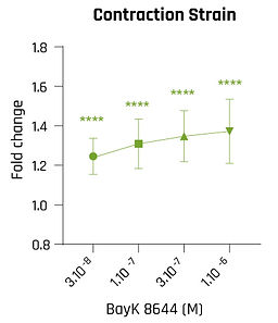
BayK 8644
Metoprolol


β-adrenergic stimulation increased beating frequency, contraction strain and contraction and relaxation speeds (N = 2; n = 16).
In this exploratory dataset, L-type Ca²⁺ channel activation increased contraction strain and contraction speed while reducing spontaneous beating frequency (N = 1; n = 11).
β1-adrenergic blockade reduced beating frequency and contraction stress in the cardiac-safety dataset (N = 2; n = 25 tissues for contractility).
Omecamtiv Mecarbil

Cardiac myosin activation increased contraction strain with little change in beating frequency and non-monotonic changes in contraction speed (N = 1; n = 10).
Istaroxime

Istaroxime increased contraction strain and both contraction and relaxation speeds, producing a luso-inotropic profile in this dataset (N = 1; n = 10).
Nifedipine

L-type Ca²⁺ channel blockade reduced contractile readouts and altered action-potential and calcium-transient parameters in the cardiac-safety dataset.
Additional pharmacological responses, experimental conditions and quantitative results are available in our scientific white papers.
FIT-FOR-PURPOSE QUALIFIED ASSAY PERFORMANCE
The SmartHeart Contractility Assay has undergone fit-for-purpose qualification for detecting and characterizing drug-induced inotropic effects in human 3D cardiac microtissues.
The qualification study evaluated seven reference compounds with distinct pharmacological mechanisms, assessing the accuracy of inotropic response detection, concentration-response relationships, assay reproducibility and analytical sensitivity.
Under the qualified experimental conditions, SmartHeart correctly identified the direction of inotropic effects for all seven reference compounds.
7/7
Correct inotropic effect detection
3
Independent experiments per compound
>20
Evaluable tissues per compound
3.7-5.7%
Minimum detectable effect by readout
Performance characterized using at least 20 evaluable cardiac rings per compound across three independent experiments. Minimum Detectable Effect values refer to the qualified study design and selected contractility readouts.
PHARMACOLOGICAL QUALIFICATION USING SEVEN REFERENCE COMPOUNDS
The assay was evaluated with five positive and two negative inotropic reference compounds targeting different components of cardiac contraction, including adrenergic receptors, calcium channels, cardiac myosin and ion transport pathways.
SmartHeart detected the expected direction of the inotropic response for all seven compounds. Concentration-response analysis enabled the estimation of EC50 or IC50 values, which were compared with available published experimental data.
These results support the use of SmartHeart for characterizing drug-induced changes in cardiac contractility within the qualified assay conditions.
Reference compound
Isoproterenol
Bay K 8644
Omecamtiv mecarbil
Istaroxime
Phenylephrine
Itraconazole
Quinidine
Inotropic effect
Positive
Positive
Positive
Positive
Positive
Negative
Negative
SmartHeart EC50/IC50
35.7 nM
24.7 nM
519 nM
161 nM
0.569 µM
914 nM
1.31 µM
ASSAY SENSITIVITY AND REPRODUCIBILITY
The SmartHeart qualification study quantified assay sensitivity through the Minimum Detectable Effect (MDE), accounting for tissue-to-tissue and experiment-to-experiment variability.
Under the qualified study design, the MDE ranged from 3.7% to 5.7% depending on the contractility readout. These values represent the smallest changes estimated to be reliably detectable with 80% statistical power at a 5% significance level, assuming an effect across approximately half of the tested concentration levels.
Reproducibility was evaluated using isoproterenol as a reference compound. After normalization to each tissue's vehicle baseline, batch-to-batch coefficients of variation remained below 10% for beating frequency and contraction strain across the four tested concentrations.
Readout
Beating frequency
Contraction strain
Mean contraction speed
Mean relaxation speed
MDE
3.7%
5.5%
4.5%
5.7%
MDE values were estimated at 80% statistical power and a 5% significance level, assuming a dose-response effect across approximately half of the tested concentrations, using at least 20 evaluable tissues from three independent experiments.
QUALIFIED ASSAY CONDITIONS AND SCOPE
The reported performance applies to SmartHeart tissues composed of human iPSC-derived cardiomyocytes and primary human ventricular fibroblasts at a 3:1 ratio, evaluated at Day 21 using acute sequential compound addition and brightfield video-based analysis.
The qualified assay measures cardiac tissue deformation (contraction strain), beating frequency and contraction/relaxation kinetics. It does not directly measure action potential duration or intracellular calcium transients.
Complementary electrophysiology, calcium-handling measurements, electrical pacing and longer exposure protocols can be incorporated according to the study objectives. These applications are distinct from the qualified core contractility workflow and may require additional characterization or qualification.
The interpretation of results depends on the tested concentration range, exposure conditions and readout-specific detection limits.
FROM CARDIAC EFFICACY TO FUNCTIONAL SAFETY
Quantitative contractility measurements support both the evaluation of intended pharmacological responses and the identification of unwanted cardiac effects.
Cardiac efficacy & pharmacological characterization
Evaluate positive or negative inotropic activity, investigate cardiac myosin and calcium-dependent mechanisms, and compare the functional responses of candidate compounds.
Functional cardiac safety
Identify unintended changes in contraction amplitude, beating activity or relaxation kinetics that may indicate functional cardiac liabilities during drug development.
ACCESS SMARTHEART CONTRACTILITY TESTING
SmartHeart is available both as a consumable platform for in-house research and through contractility testing services performed by 4Dcell.
Run experiments in your laboratory
Use SmartHeart® 96-well plates to generate human 3D cardiac microtissues and investigate drug-induced contractile responses with your own experimental workflow.
The glass-bottom format supports brightfield imaging, while compatible SmartX analysis workflows enable quantitative assessment of tissue contractility.
Let 4Dcell perform your study
Access our fit-for-purpose qualified contractility testing service, from experimental design and compound exposure to quantitative analysis and scientific reporting.
Our team can adapt study designs to your compound's mechanism of action and research objectives, with additional assessment where required.