
Cardiac spheroids vs organoids: a guide to 3D heart models
State of the art of innovative cardiac 3D cell culture systems
December 2023
What is the difference between cardiac spheroids and cardiac organoids?
Cardiac spheroids and cardiac organoids are both three-dimensional cellular models, but they differ primarily in how they are generated and the degree of tissue organization they reproduce.
Cardiac spheroids are generally formed by aggregating pre-differentiated cardiac cells, such as human induced pluripotent stem cell-derived cardiomyocytes (hiPSC-CMs), cardiac fibroblasts and endothelial cells. Their cellular composition can be defined before assembly, making them useful for studying cell interactions and functional responses under controlled experimental conditions.
Cardiac organoids are typically generated from pluripotent stem cells or cardiac progenitors that differentiate and self-organize into multicellular structures. Depending on the protocol, they can reproduce aspects of cardiac morphogenesis, cellular diversity and early heart development.
Engineered heart tissues (EHTs) represent another category of 3D cardiac models. They use bioengineering approaches to control tissue geometry, attachment, mechanical loading or extracellular matrix organization. These properties can facilitate quantitative measurements of cardiac contraction and relaxation.
The terminology is not completely standardized, and some experimental systems combine features of multiple categories. Therefore, the method of generation, tissue architecture and validated functional readouts are more informative than the model name alone. [1, 12]
How are human cardiac organoids generated?
Human cardiac organoids are commonly generated by differentiating pluripotent stem cells, including human iPSCs, through signaling pathways involved in early cardiovascular development.
Many protocols rely on the timed modulation of WNT signaling to guide cells through mesoderm specification, cardiac progenitor formation and subsequent cardiac differentiation.
Under suitable conditions, differentiating cells can self-organize into three-dimensional structures containing multiple cardiovascular cell populations, including cardiomyocytes and, depending on the model, endothelial, endocardial, epicardial or smooth muscle-like cells. [2–4]
An important development in the field is the generation of chamber-specific cardiac organoids, also known as cardioids. Schmidt et al. (2023) demonstrated a multi-chamber cardioid platform representing major embryonic heart compartments, including atrial and ventricular regions. The study provided an experimental system for investigating chamber interactions and developmental defects. [5]
These approaches are particularly relevant for studying cardiac morphogenesis, congenital heart disease and developmental responses to genetic or pharmacological perturbations.
However, cardiac organoids generally reproduce selected developmental features rather than the complete structure or physiology of an adult human heart.

Figure 1: Stages of differentiation of cardiac organoids[3] using a protocol developed by Drakhlis et al.[4] Figure adapted from Kim et al.[3]
How are cardiac spheroids generated, and why does cell composition matter?
Cardiac spheroids are generally produced by bringing differentiated cardiac cells together under culture conditions that promote cell–cell adhesion and three-dimensional aggregation.
Human iPSC-derived cardiomyocytes are frequently combined with other cardiac cell populations to reproduce selected features of the myocardial microenvironment.
The choice of cell types depends on the experimental objective:
-
Cardiomyocytes provide the contractile activity of the model.
-
Cardiac fibroblasts contribute to extracellular matrix production, structural organization and intercellular signaling.
-
Endothelial cells enable the investigation of vascular-related signaling and endothelial–myocardial interactions.
-
Smooth muscle cells can contribute to multicellular models investigating vascular or tissue-specific interactions.
Kahn-Krell et al. (2022) investigated cardiac spheroids combining cardiomyocytes, endothelial cells, smooth muscle cells and fibroblasts derived from human iPSCs, reporting functional differences associated with multicellular composition. [7]
Similarly, Polonchuk et al. (2017) demonstrated that multicellular cardiac spheroids could reproduce selected structural and cellular characteristics of the human myocardial microenvironment, including extracellular matrix organization and vascular-like structures. [8]
These findings highlight the importance of defining cell composition according to the biological question rather than assuming that a larger number of cell types automatically produces a better model.

Figure 2: Formation of cardiac spheroids from human primary cells (left) and cells differentiated from iPSCs (right). Figure adapted from Polonchuk et al.[8]
Can cardiac spheroids and organoids model human heart diseases?
Three-dimensional cardiac models can be adapted to investigate specific cardiovascular disease mechanisms by selecting relevant cell populations, genetic backgrounds or experimental conditions.
For example, cardiac spheroids can be used to study myocardial fibrosis through interactions between cardiomyocytes, fibroblasts and extracellular matrix components.
Figtree et al. (2017) investigated a cardiac fibrosis model using spheroids derived from neonatal rat cardiac cells and exposed to transforming growth factor beta (TGF-β), a profibrotic signaling molecule. The study demonstrated changes in extracellular matrix-associated characteristics under profibrotic stimulation. [9]
Human iPSC-based cardiac models offer additional opportunities to investigate inherited cardiomyopathies and disease-associated genetic variants.
The suitability of any model depends on the disease mechanism being investigated, the cellular phenotype reproduced and the availability of relevant functional measurements.
Importantly, a model that reproduces one disease-associated phenotype should not automatically be considered representative of the entire human disease.
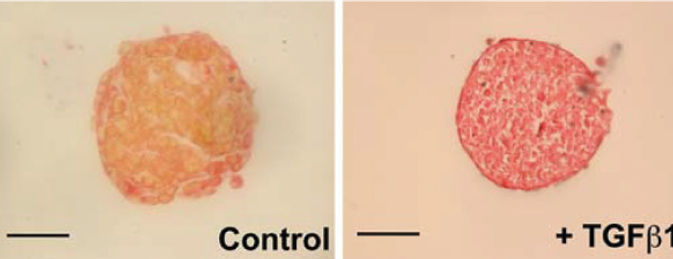
Figure 3: Neonatal rat cardiac spheroids- Staining of the muscular (yellow) and ECM (red) components of cardiac spheroids. The predominant red stain with the profibrotic agent TGF-beta demonstrates the creation of a fibrosis disease model. Scale bar, 100 μm.Figure adapted from Figtree et al.[9]
What methods are used to produce and culture cardiac spheroids and organoids?
Several culture methods are available for generating three-dimensional cardiac cell aggregates. Their suitability depends on the required tissue size, cellular composition, reproducibility, imaging conditions and experimental throughput.
Hanging-drop culture
Cells are suspended in small droplets, allowing gravity and cell–cell adhesion to promote aggregation. This approach is useful for spheroid formation but can require careful handling during prolonged culture.
Ultra-low-attachment plates
Non-adherent culture surfaces encourage cells to aggregate rather than attach to the culture vessel. These systems are widely used for 3D cell culture, although aggregate uniformity and positioning depend on the specific plate design and protocol.
Microwell-based culture
Microwell arrays provide defined locations for cell aggregation and can help control spheroid size through the geometry of the microwells and the initial cell seeding density. Multiwell plate integration can facilitate parallel experiments and imaging.
Dynamic suspension systems
Spinner flasks and other suspension culture approaches can support the generation and maintenance of larger numbers of aggregates. However, shear stress, aggregate size distribution and handling conditions must be controlled.
Each method involves trade-offs between biological complexity, reproducibility, manipulation requirements and compatibility with downstream analysis. [10, 12]
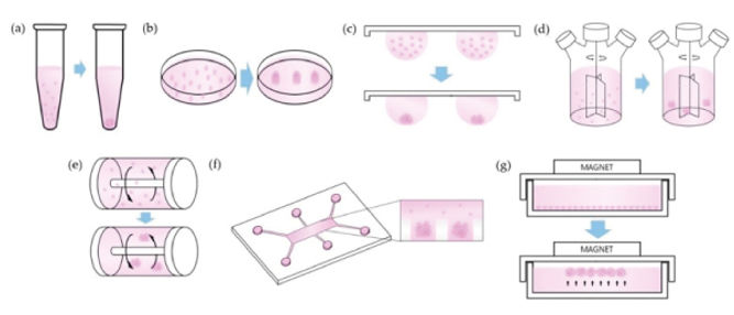
Figure 4: Techniques used to keep cell aggregates in suspension for spheroid tissue culture. Figure adapted from Ryu et al.[10]
How can micropatterning guide cardiac tissue organization?
Micropatterning provides an alternative approach to controlling cardiac cell organization by defining where cells can attach and grow.
By restricting cell adhesion to specific geometries, such as disks, lines or other patterns, researchers can investigate how spatial confinement and mechanical cues influence cell differentiation and tissue organization.
Ma et al. (2015) demonstrated that geometrically confined human pluripotent stem cell colonies could self-organize into beating cardiac microchambers. The study showed that spatial constraints, combined with modulation of WNT signaling, influenced cardiac lineage specification and tissue morphogenesis. [11]
These findings illustrate the importance of physical microenvironmental cues in cardiac development.
Micropatterning can therefore complement conventional spheroid and organoid culture by introducing spatial control over cell organization, particularly in studies of differentiation, morphogenesis and cell–cell interactions.
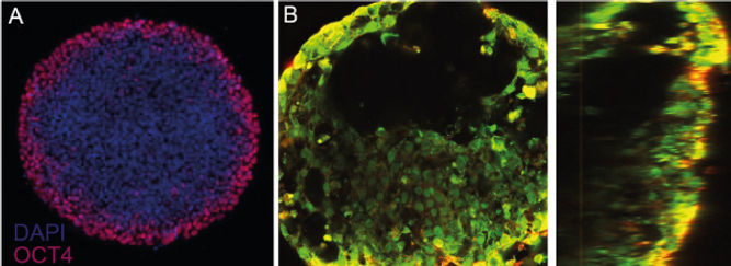
Figure 5: Micropatterned organoids. A) Loss of OCT4 expression at the periphery of the micropattern indicates loss of pluripotency. (Disk-shaped micropattern 600 um in diameter.) B) Following differentiation protocols, microchambers devoid of cells are visible in top (left) and side (right) views. Figure adapted from Ma et al.[11]
Cardiac spheroids vs organoids vs engineered heart tissues: which model should you choose?
The choice of a 3D cardiac model should be guided by the biological question and the measurements required rather than by model complexity alone.
Cardiac spheroids — for controlled multicellular studies
Cardiac spheroids are particularly useful when researchers need to investigate interactions between defined cardiac cell populations, examine tissue responses to compounds or develop reproducible multicellular assays.
Cardiac organoids — for developmental biology and complex tissue organization
Cardiac organoids are well suited to studying aspects of human cardiac differentiation, morphogenesis, congenital defects and the emergence of multicellular structures.
Engineered heart tissues — for quantitative cardiac function
Engineered cardiac tissues can provide controlled geometries and mechanical boundary conditions. Depending on the platform, they enable measurements of tissue contraction, relaxation kinetics, force-related parameters and responses to pharmacological stimulation.
Micropatterned cardiac models — for spatially controlled differentiation
Micropatterned systems enable researchers to investigate how geometric confinement influences cell fate, organization and tissue formation.
These approaches are complementary. No single 3D cardiac model reproduces every aspect of human heart physiology or provides every functional readout required for preclinical drug testing.
What functional readouts are important for cardiac drug testing?
Three-dimensional cardiac models can provide valuable information about drug-induced changes in cardiac function, provided that the model and measurement technology are suitable for the intended application.
Important functional readouts include:
-
Contractility: contraction amplitude, tissue deformation, contraction strain and, where supported by the measurement configuration, force-related parameters.
-
Contraction and relaxation kinetics: measurements of how rapidly tissues contract and relax, which can help characterize inotropic and lusitropic responses.
-
Beating frequency and regularity: changes in spontaneous activity, beat rate and rhythm stability.
-
Calcium handling: intracellular calcium transient amplitude, frequency and kinetic properties.
-
Electrophysiology: action potential characteristics, repolarization and rhythm-related responses, using appropriate recording methods.
Not all cardiac spheroids or organoids provide these measurements directly. Some readouts require specialized imaging, mechanical supports, electrical stimulation or complementary assays.
Consequently, the ability to obtain robust, quantitative functional data is a critical criterion when selecting a 3D cardiac model for safety pharmacology or drug efficacy assessment.
What are the limitations of 3D cardiac spheroids and organoids?
Although 3D cardiac models provide important advantages over conventional monolayer cultures, their limitations must be considered when designing experiments and interpreting results.
Cellular maturity
Human iPSC-derived cardiomyocytes often retain features of immature cardiac cells. Three-dimensional organization alone does not guarantee adult-like electrophysiological, metabolic or mechanical properties.
Tissue composition
The absence or overrepresentation of specific cell populations can affect the relevance of a model for a particular biological question.
Diffusion and tissue size
Oxygen, nutrient and compound diffusion can vary within large cellular aggregates, potentially creating heterogeneous microenvironments.
Experimental variability
Differences in initial cell number, aggregate size, differentiation efficiency or culture conditions can influence tissue characteristics and downstream responses.
Functional accessibility
Some 3D models are difficult to position, stimulate or image consistently. Quantitative measurements of contractility or electrophysiology may require specific adaptations.
Physiological scope
Even advanced models reproduce only selected aspects of the human heart. Their predictive relevance must be evaluated against the intended context of use.
Addressing these limitations is essential when translating exploratory 3D culture methods into reproducible preclinical testing workflows.
Improving 3D culture reproducibility with anchored spheroids
For experimental workflows involving repeated measurements, medium exchanges or compound additions, spheroid stability and positioning can become important practical constraints.
Conventional non-adherent spheroids may move during liquid handling, making it more difficult to track the same aggregate over time or maintain consistent imaging conditions.
4Dcell developed SmartSphero™, a hydrogel-based microwell platform incorporating proprietary Anchorage Point Technology (APT®).
The platform is designed to support the formation of reproducible 3D cell aggregates while keeping them anchored at defined positions.
Its key features include:
-
Defined microwell geometry supporting spheroid formation.
-
Localized anchoring to reduce displacement during routine handling.
-
Predictable spheroid positioning for repeated imaging.
-
Multiple aggregates per well for parallel measurements.
-
Integration with standard multiwell plate formats.
These design principles address several practical challenges associated with conventional spheroid culture and can support more structured workflows for 3D cell model development and screening.
Explore the SmartSphero™ platform to learn more about anchored spheroid culture.
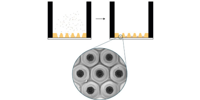
Figure 6: 4Dcell SmartSphero plates create homogenous spheroids, anchored at a small (~100 um) contact point, that are not lost during medium exchanges
Beyond cardiac spheroids: engineered human 3D cardiac microtissues for functional testing
While conventional spheroids and developmental organoids provide valuable biological models, preclinical cardiac drug testing often requires reproducible tissue organization and quantitative functional measurements.
Engineered cardiac microtissues can address some of these requirements by combining three-dimensional cellular organization with controlled geometry and mechanical loading.
4Dcell's SmartHeart® platform uses human iPSC-derived cardiomyocytes and cardiac fibroblasts that self-assemble into ring-shaped 3D microtissues around central hydrogel pillars.
The platform incorporates nine cardiac microtissues per well in a standard 96-well plate format, enabling multiple tissue-level observations under the same experimental condition.
Tissue contractility can be quantified through brightfield video analysis, including contraction strain, beating frequency and contraction/relaxation kinetics.
Complementary electrophysiological and calcium-handling measurements can provide additional mechanistic information depending on the study design.
This approach illustrates how engineered cardiac geometry can extend the experimental capabilities of conventional 3D cell aggregates.
Learn more about SmartHeart® and its applications in cardiac drug testing.
Conclusion: selecting the right human 3D cardiac model
Cardiac spheroids, organoids and engineered heart tissues provide complementary approaches to investigating human cardiac biology.
Cardiac spheroids enable controlled studies of multicellular interactions, while developmental organoids provide insights into cardiac morphogenesis and tissue organization. Engineered heart tissues introduce additional control over mechanical conditions and functional measurement.
For drug discovery and preclinical testing, the most appropriate model is not necessarily the most structurally complex one. Model selection should prioritize biological relevance, reproducibility, validated readouts and compatibility with the intended experimental workflow.
Advances in cell engineering, tissue organization and quantitative analysis are helping researchers translate 3D cardiac models into increasingly accessible experimental systems.
Explore 4Dcell's human 3D cardiac platforms or discuss your experimental requirements with our scientific team.
References
[1] Simunovic, M. & Brivanlou, A. H. Embryoids, organoids and gastruloids: new approaches to understanding embryogenesis. Development 144, 976–985 (2017).
https://doi.org/10.1242/dev.143529
[2] Ramirez-Calderon, G., Colombo, G., Hernandez-Bautista, C. A., Astro, V. & Adamo, A. Heart in a Dish: From Traditional 2D Differentiation Protocols to Cardiac Organoids. Frontiers in Cell and Developmental Biology 10, 855966 (2022).
https://doi.org/10.3389/fcell.2022.855966
[3] Kim, H., Kamm, R. D., Vunjak-Novakovic, G. & Wu, J. C. Progress in multicellular human cardiac organoids for clinical applications. Cell Stem Cell 29, 503–514 (2022).
https://doi.org/10.1016/j.stem.2022.03.012
[4] Drakhlis, L. et al. Human heart-forming organoids recapitulate early heart and foregut development. Nature Biotechnology 39, 737–746 (2021).
https://doi.org/10.1038/s41587-021-00815-9
[5] Schmidt, C. et al. Multi-chamber cardioids unravel human heart development and cardiac defects. Cell 186, 5587–5605.e27 (2023).
https://doi.org/10.1016/j.cell.2023.10.030
[6] Yang, X., Cheng, K., Wang, L.-Y. & Jiang, J.-G. The role of endothelial cell in cardiac hypertrophy: Focusing on angiogenesis and intercellular crosstalk. Biomedicine & Pharmacotherapy 163, 114799 (2023).
https://doi.org/10.1016/j.biopha.2023.114799
[7] Kahn-Krell, A. et al. A three-dimensional culture system for generating cardiac spheroids composed of cardiomyocytes, endothelial cells, smooth-muscle cells, and cardiac fibroblasts derived from human induced-pluripotent stem cells. Frontiers in Bioengineering and Biotechnology 10, 908848 (2022).
https://doi.org/10.3389/fbioe.2022.908848
[8] Polonchuk, L. et al. Cardiac spheroids as promising in vitro models to study the human heart microenvironment. Scientific Reports 7, 7005 (2017).
https://doi.org/10.1038/s41598-017-06385-8
[9] Figtree, G. A., Bubb, K. J., Tang, O., Kizana, E. & Gentile, C. Vascularized Cardiac Spheroids as Novel 3D in vitro Models to Study Cardiac Fibrosis. Cells Tissues Organs 204, 191–198 (2017).
https://doi.org/10.1159/000477436
[10] Ryu, N.-E., Lee, S.-H. & Park, H. Spheroid Culture System Methods and Applications for Mesenchymal Stem Cells. Cells 8, 1620 (2019).
https://doi.org/10.3390/cells8121620
[11] Ma, Z. et al. Self-organizing human cardiac microchambers mediated by geometric confinement. Nature Communications 6, 7413 (2015).
https://doi.org/10.1038/ncomms8413
[12] Idais, D., Roche, C. D., Kalogianni, G., Polonchuk, L. & Gentile, C. Generation and applications of cardiac spheroids. npj Biomedical Innovations 2, 17 (2025).
https://doi.org/10.1038/s44385-025-00024-y